Sandfish and teeth: the power of bionics
UV-2600 and ISR-2600Plus: analysis of nanospikes from TiO2 and sodium on surfaces from dental materials
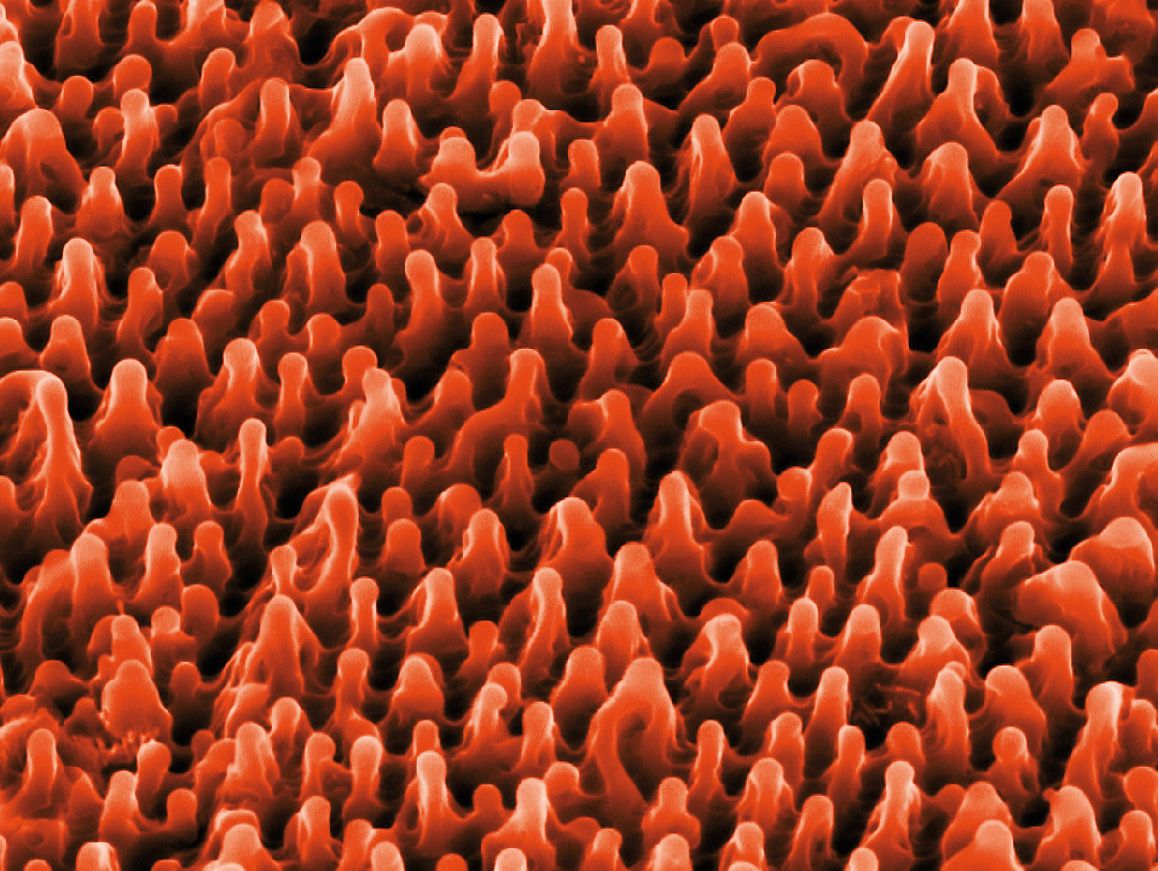 Figure 1: Nanospiking on surface
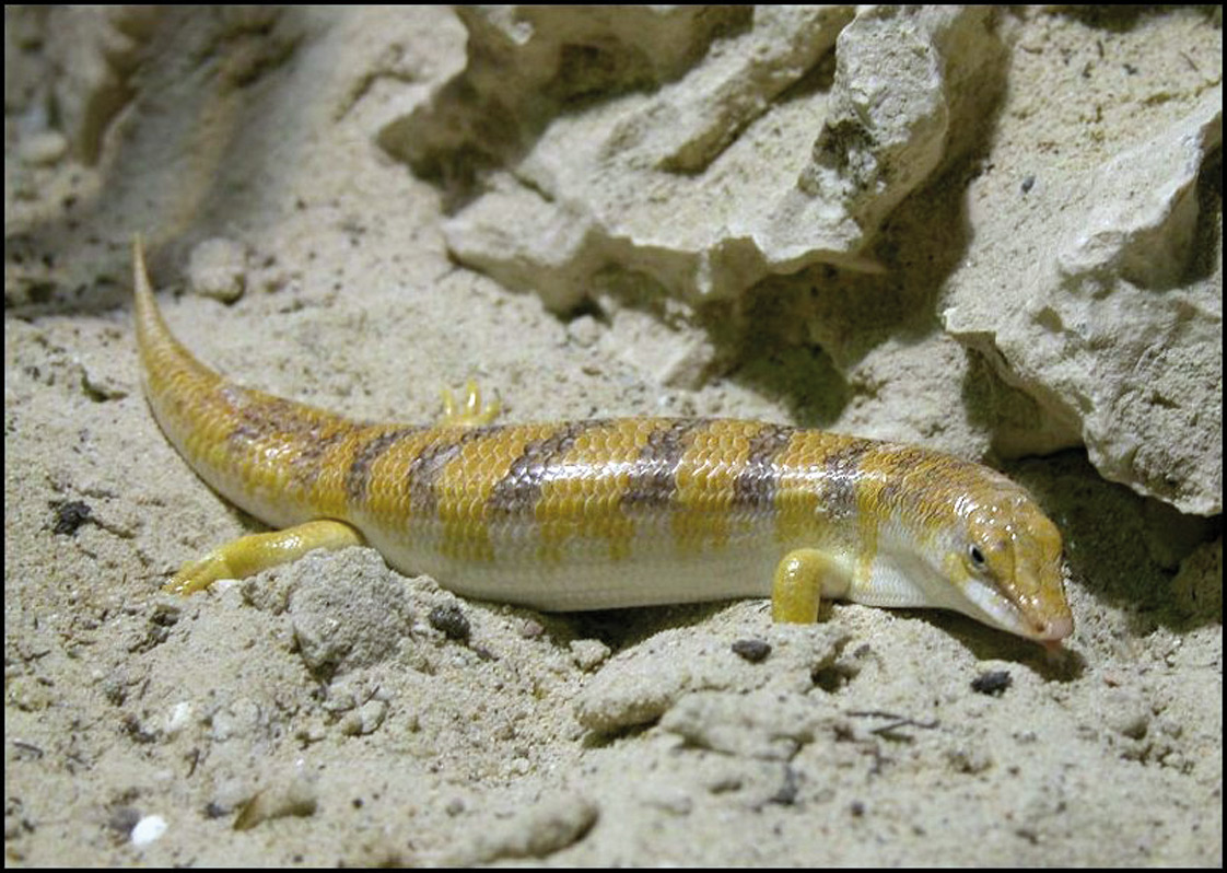 Desert Sandfish (Scincus scincus)
Foto: Wilfried Berns Tiermotive.de
What has a reptile that swims through sand got in common with paint? Both are linked through bionics, an interdisciplinary science which applies biological methods and systems found in nature to design modern technologies. A well-known application is the so-called lotus effect imitating the nanoscopic properties of plants for coatings, paints, and roof tiles to support self-cleaning features.
Paint whiteners and dental implants based on Titanium might seem a world away from the world of the desert sandfish (Scincus scincus), a little reptile that swims through sand like through water. But advances in the world of bionics makes the reptile’s skin and the surface of the dental implants share the same secret: nanospikes.
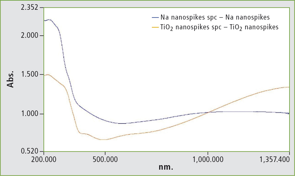 Figure 2: UV-VIS-NIR spectra from nanospikes (Na dark blue line, Titanium Dioxide orange line)
The skin of the desert sandfish has evolved spiky nanostructures which produce a virtually frictionless surface, offering only a tiny fraction of the normal surface area of the creature in contact with the hard grains of dry sand. [1] In the dental implant world, the nanospikes exploit almost the exact opposite of this property, providing a very large contact area for the implant to establish contact with the soft surface of the tissues.
This application discusses the analysis of TiO2-based nanospike material for dental purposes.
New approach: nanostructures on surfaces related to or with TiO2
For decades, TiO2 has been a well known substance for different areas of application. Very typical is its use as a whitener, based on excellent reflectance properties. Nanospiking opens up additional applications for TiO2. Nanospiking is an approach generating thin active surfaces with TiO2 to get the antibacterial properties of the TiO2 into very small areas. Nanospiking also produces a high surface reflectance composed of many tens of thousands of tiny spikes. The illustration on page 6 depicts titanium nanospikes at high magnification using Scanning Electron Microscopy (SEM) (figure 1). This spiking gives the surface a very high intense and bright white appearance.
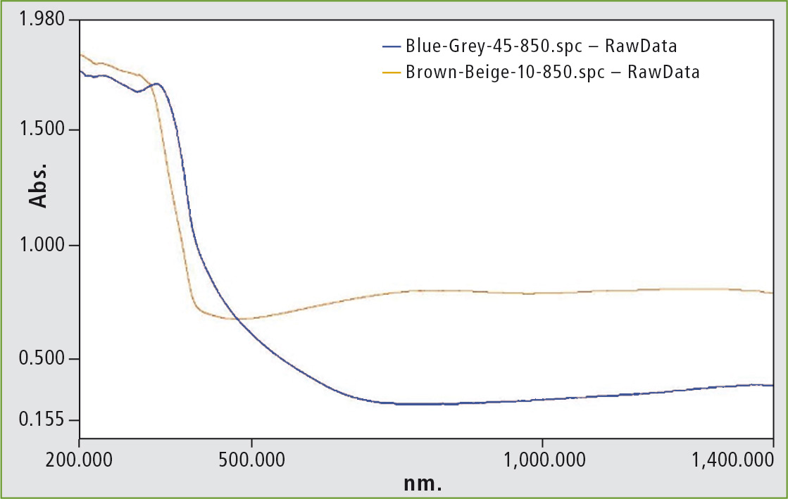 Figure 3: Brown-beige material (paler trace) and blue-grey material (darker trace)
However, the unusual surface structure when presented in the nanospiked form can alter the reflectivity. Titanium Dioxide also exhibits fluorescence. For medical use, it is usually advantageous to use as little material as possible. A new approach is to use nanostructures on surfaces related to or with TiO2. The nature of the nanospikes represents that of a field of needles which, in principal, is an ideal diffuse reflector. Hence, the expectation is that such surface has a higher diffuse reflectance level than a normal un-spiked surface.
Application
Several samples were measured with UV-VIS-NIR spectroscopy in reflectance mode. [2, 3] There were Na and TiO2 nanospiked samples, one blue-grey and one brown-beige shiny material. Due to the nature of the material, an integrating sphere was used to collect as much diffuse reflectance as possible. These samples were small in size, so in order to mount the samples on the reflectance port of the ISR-2600Plus integrating sphere, a background mount was made with BaSO4 white standard. The sample was large enough for the beam image of the instrument to fall only onto the sample.
The sample was mounted on a white reflector, so that the reflection losses from the sphere were minimized. Scans were made from 220 to 1,400 nm using the unique dual detector integrating sphere with the sample mounted at the diffuse reflectance port, which is at a zero degree incident angle to reject any specular reflectance components of the measurement.
Summary
The nanospike traces show high absorbance (low reflection) at short wavelengths; it is likely that the trace at 200 nm is limited by the reflectivity of the inside coating of the sphere: (Barium sulfate) which mainly falls in the region between 220 and 200 nm. Even in this region however, the results show that the instrument was still operating within range as the data remains smooth. If the limit of detection had been reached, the trace would have become ‘noisy’ in the region below 220 nm.
The remainders of the traces are well within the photometric range and the stray light range of the system. The reflectance traces are very clear with minimal noise. These traces are recorded in ‘normal’ specular excluded mode, so the specular reflectance of the sample is rejected. (The sample is illuminated at near-normal angle and mounted in a way that specular reflectance is lost through the sphere transmission port.)
With materials which do not reflect an image at anything but very shallow viewing angles, this measurement is generally the most accurate as the specular component is minimal. In the event that the sample is somewhat specular, an additional measurement can be made using the ‘specular included’ reference port which illuminates the sample at eight degrees from normal, allowing specular reflectance to be reflected back to the sphere wall and thus included in the measurement.
Reviewing these results, the instrument clearly delivers accurate results for the entire 200-1,400 nm range. The results are all obtained using barium sulfate as a reference material. This is a very good neutral reflector. All Integrating Sphere measurements have to be made in Percent of Reference mode as there is no way of measuring diffuse reflectance as an absolute value. [2, 3] The instrument normalizes all the reflectance values for the barium sulfate to 100 % (0 Abs) during background acquisition, but consideration should be given to the reflectivity of the reference material.
Literature
[1] The Sandfish of the Sahara – A Model for Friction and Wear Reduction, Ingo Rechenberg, Abdullah Regabi El Khyari, Technische Universität Berlin, Bionik und Evolutionstechnik, 2/2004.
[2] Standards in Absorption Spectrometry, Burgess and Knowles (ISBN 0-412-22470-4).
[3] Fundamentals of Molecular Spectroscopy, Banwell and McCash (ISBN 0-07-707976-0).
Further information on this article
Application note: Nanospikes and sandfish