Life can be hard
Contamination of bread – Particle analysis with FTIR-ATR single reflection ATR technique and EDX for inorganic analysis

With a bright smile, the author has bought healthy full grain bread, persuaded to do something good for the body. Being hungry, he ate the bread straight away and maybe a little too fast. Suddenly – a cracking sound, he has bitten on something incredibly hard. Oh no, the teeth! Is one broken? Repair is so expensive, it would be a disaster. Quickly checked, everything okay. But a hard particle needs investigating…
Once isolated, the particle looked like a rice corn which had dried to be hard as a stone.
Let’s name this particle (figure 1). It has a size of 3 to 5 mm and a shape like a rice corn. Under the stereo microscope, it showed an orange color. Under normal day light, it appeared white. Having being hurt by this particle, it was of interest to analyze the real chemical consistence.
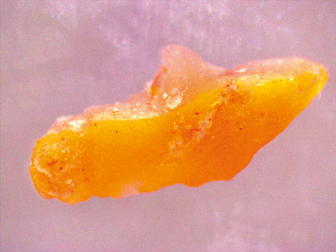 Figure 1: The contaminant in wholegrain bread has a size of 2 x 5 mm; this photo was obtained with a camera integrated in a stereo microscope
Particle measurement techniques
For a rough screening and identification of materials, two non-destructive analysis techniques are available: EDX and FTIR. Non-destructive means that the sample keeps its form and that a chemical treatment is not needed.
EDX (energy dispersive X-Ray fluorescence spectrometer) makes the analysis easy due to the “place and measure” nature of its application. Infrared spectroscopy (FTIR Fourier Transform Infrared Spectroscopy) is a measurement technique which helps to identify very hard material using the single reflection ATR (Attenuated Total Reflection) tool without any other physical treatment. This is possible because the ATR unit Specac QuestTM has an integrated diamond window which makes it possible to analyze very hard solid materials at their surfaces (penetration depth of the infrared beam of ~2 µm). The hardness of the diamond will help to do so.
Sample preparation and spectral analysis using FTIR
No special sample pretreatment was used. The particle had a rice-corn shape of 3 by 5 mm. It was simply placed on the diamond window of approx. 2 mm size and the anvil (accessory) was screwed down to press the corn onto the diamond window, breaking it into smaller, still very hard pieces. At this moment, it became obvious that an inorganic material was present. FTIR measurement was done in a range of 400 to 4,600 cm-1. Infrared reflection spectra are presented in figures 2 to 4.
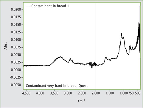 Figure 2: Low-pressure: infrared spectrum from the surface of the particle, which is dominated by cellulose
Anvil pressure was set in three levels. With low pressure a cellulose spectrum was measured at the effective top surface. This is acceptable because the environment of this specific corn was previously bread. Traces of cellulose remained on the surface of the particle. Figures 3 and 4 show measurements of fragments from the corn and of the crystal dust. The dust appeared after the particle cracked and was collected from the measurement crystal. Both measurements with different pressure – middle (fragment) and high pressure (particle dust) resulted in an inorganic spectrum. The spectrum is most probably the SiO2 spectrum. A library search resulted in a good match dedicated to diatomaceous earth.
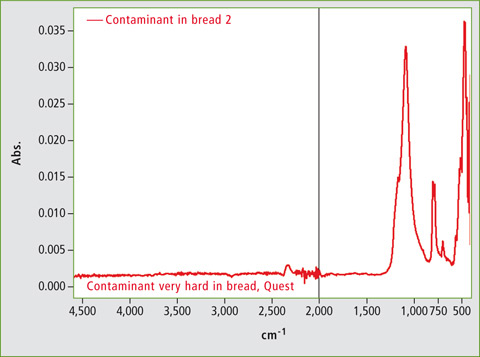 Figure 3: Middle- pressure: infrared spectrum from a collection of particles from the corn, mostly identified as SiO2
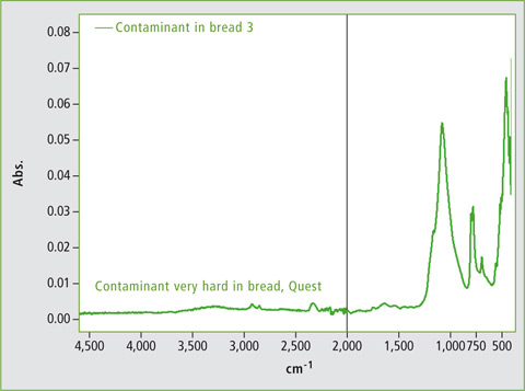 Figure 4: high-pressure: reconfirming the result from 2, a collection of fine crystals from the particle separated powder
Sample preparation and X-ray analysis with EDX-8000
In addition to FTIR-ATR measurements, EDX technique was applied to obtain elemental composition of the sample in the ppm to percentage level without the need of sample preparation. Standards for calibrating the instrument are useful to increase accuracy of measurement results, but are not mandatory. The analysis is non-destructive, making the technique very useful for screening of unknown samples.
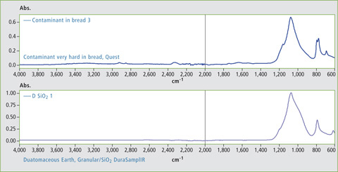 Figure 5: Library search and hit with diatomaceous earth, granular/SiO2 DuraSampllR
For this measurement, the particle was transferred to a special plastic cup. With a collimator (standard accessory) the measurement range was narrowed down to 1 mm. Using a camera (standard accessory), the exact position of the sample within the sample chamber was confirmed. For the measurement, a simple standardless FP (fundamental parameter) method was used. Calibration of the instrument was not needed. No filters (standard accessory) or vacuum condition (optional accessory for measurement of light elements) were used.
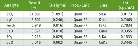 Table 1: EDX results using Fundamental Parameter (FP method)
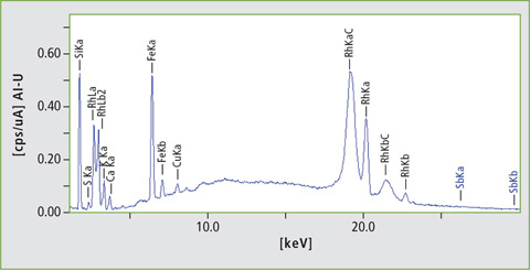 Figure 6: EDX spectrum of the particle
Results
It was clear that the main compound of the sample was SiO2, i.e. a stone. Other elements present such as Fe are typical elements occurring in stones and minerals.
Instrumentation
- IRTracer-100 with LabSolutions IR software
- Single reflection unit – Specac Quest™
- Shimadzu Libraries
- EDX-8000
- EDXIR Contaminant Library
Conclusion
Infrared spectroscopy can identify the major components in natural and complex sample material. The domain of infrared is the identification of substances from organic or inorganic characteristics. As inorganics have more unspecific signal groups, EDX is a good complementary analysis for the elemental distribution in a small particle such as the corn in this case.