A bit like lasagna:
Proper slicing and analysis show what packaging film is really made of
Microtome sections reveal their secrets with the AIRsightTM infrared and Raman microscope


Dr. Kai Klein, Dr. Aikaterini Karatzia, Shimadzu Europe
Modern plastic film packaging used in the food and pharmaceutical industries keeps products fresh and protected. This packaging typically consists of multiple layers bonded together, and analyzing these layers is important for quality control, recycling and more. However, doing that can be quite complicated. How do you measure multiple layers of a film that itself is only a few micrometers thick? One new way to do that is to apply the advantages of distinct instruments in a novel way.
Consider lasagna
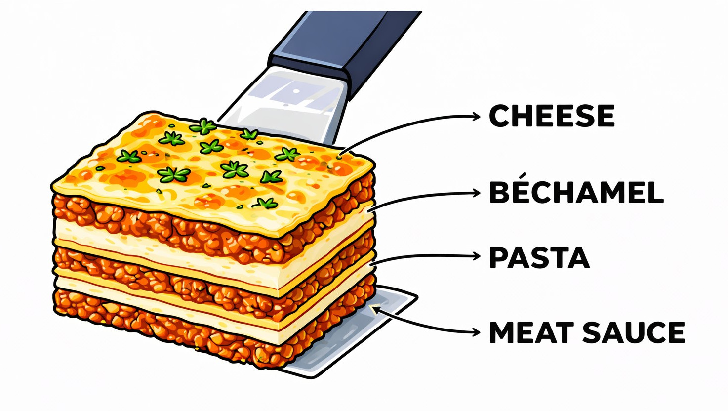
It’s time for dinner, and you’re hungry. Tonight, you’re looking forward to one of your favorite dishes: lasagna. Suddenly, it’s there on your plate. You have a good overview of what it looks like, and it does look good! Maybe you are so hungry or so eager to enjoy the experience of lasagna in your mouth that you immediately start eating. Or, perhaps, you pause for a moment to further inspect the finer points of your dinner. You gently slice through your little piece of heaven and inspect its inner workings. The cross-section reveals all: layers of meat sauce, béchamel, pasta and cheese which together create a culinary sensation (Figure 1).
In a similar way, it takes many different layers of plastic film to create a fully functional piece of modern packaging. Modern packaging, such as plastic films used in the food and pharmaceutical industries, keeps products fresh and durable, protects their contents and usually saves weight compared to similarly effective alternatives. These packages typically consist of multiple layers bonded together, which can include different materials depending on their intended application. Commonly, there is a surface layer acting as a barrier against oxygen and/or moisture, a carrier layer providing stability and a printed layer designed to appear attractive to the customer. In rarer cases, an additional layer may be included to protect against odor or light.
Analyzing these layers is of great importance for a number of reasons: for scientific research, material development, quality control, in waste management and recycling. For disposal and recycling, for example, it is essential to know exactly which material is to be recycled. And, from a scientific perspective, this knowledge makes it possible to continuously improve and refine packaging.
Slicing: The first challenge of analyzing plastic film packaging
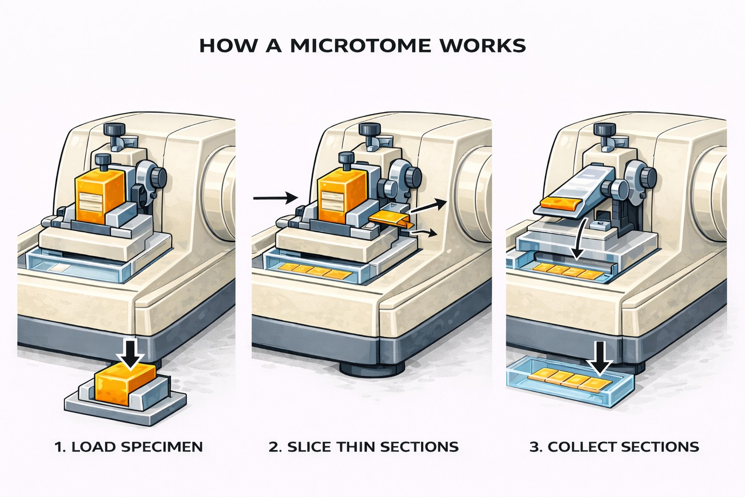
However, analyzing these layers can sometimes be quite complicated. How do you measure multiple layers of a film that itself is only a few micrometers thick? To understand why packaging works so well, it is worth taking a look into this microscopic world. That’s where the microtome cutting tool comes into play.
With the help of a microtome, extremely thin and smooth slices of these materials can be produced. The cross-section obtained with the microtome provides insights into the number, sequence and thickness of individual layers, the material transitions and adhesive layers, as well as any irregularities or defects that could lead to reduced shelf life. Therefore, regular analysis of packaging materials in the food and pharmaceutical sectors is of great importance for quality assurance.

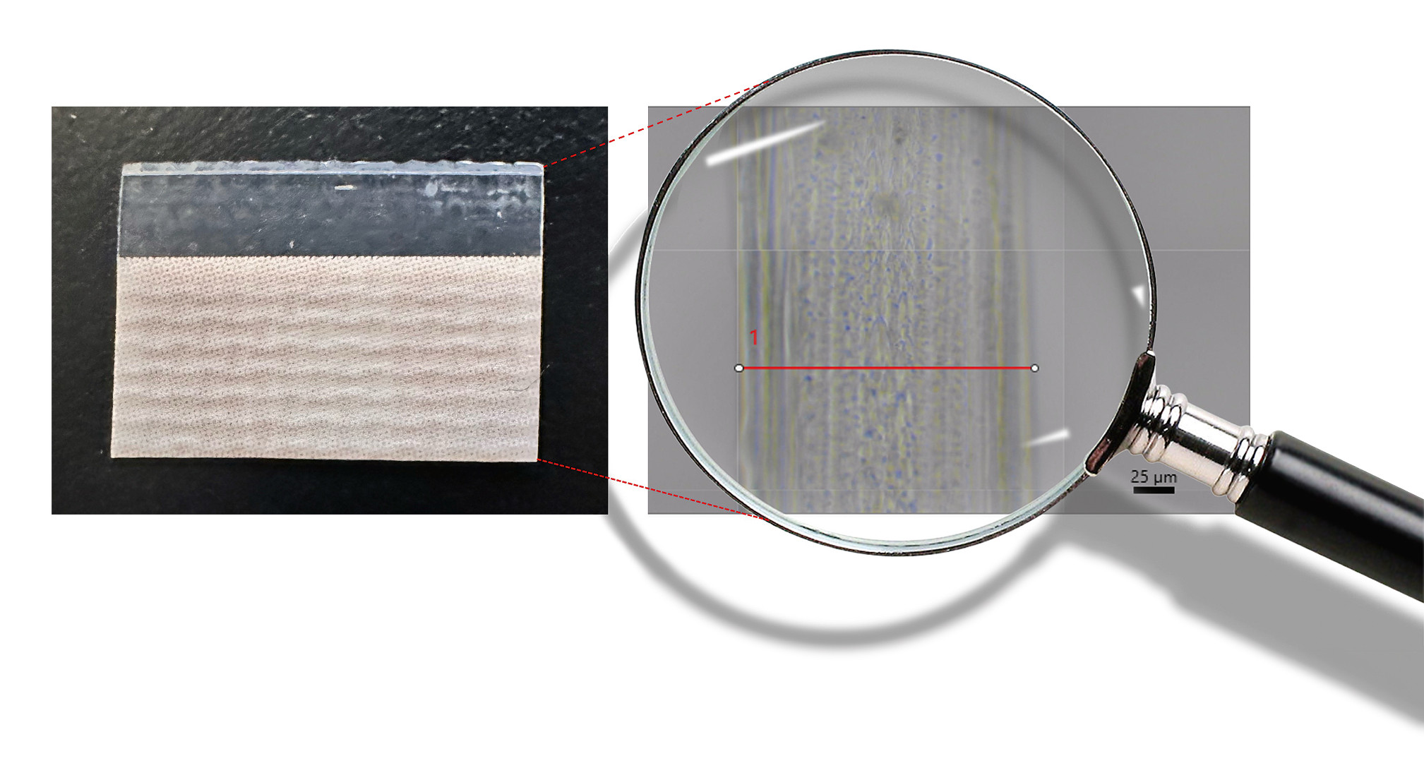
After selecting a suitable piece of the sample, it is clamped into the microtome holder. For multilayer films, it is recommended to position the sample perpendicular to the cutting direction to ensure a clean cross-section (Figure 2). Depending on the type and stability of the sample, it is either clamped directly or embedded beforehand into a suitable stabilizing medium, such as synthetic resin. For embedding very thin films, it is advisable to choose a medium that is chemically inert to the material being examined and does not require excessive heat.
The choice of blade and the thickness of the cut layer often depend on the planned analysis. For transmission measurements with an infrared microscope, thinner sections are cut using harder blades compared to, for example, reflection measurements. The cutting process itself should be carried out in a smooth and steady motion to avoid unwanted rough spots on the surface, which could distort or complicate later analysis under the microscope. The separated thin sections are then prepared according to the chosen analytical method. For transmission measurements, the sections can be lightly fixed at the ends with adhesive tape so that the area to be examined remains exposed. For reflection measurements, the sections can be placed on a metal-coated surface or, if sufficiently thick, on a standard glass microscope slide.
How a mictotome works
Analyzing: The second challenge of analyzing plastic film packaging
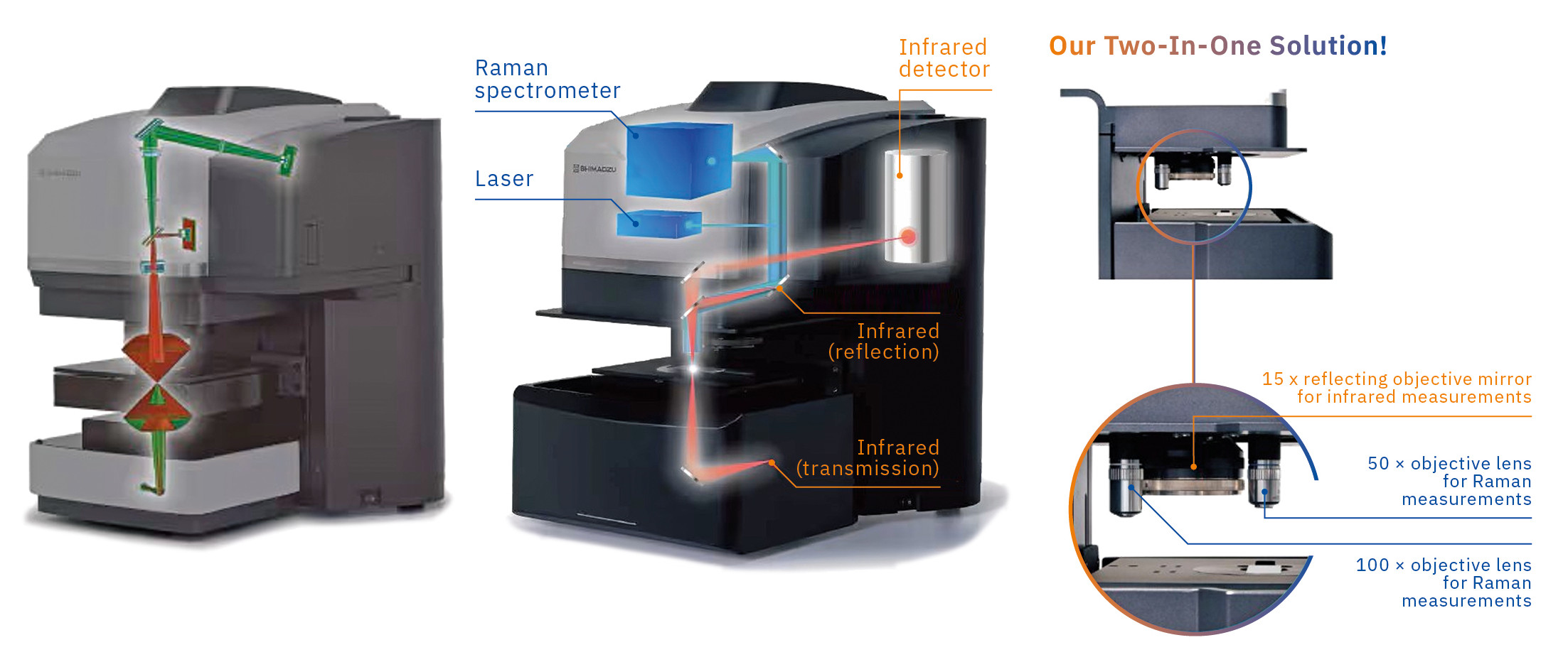
Infrared microscopy enables the acquisition of infrared spectra at defined locations on a sample surface. The AIMsight™ and AIRsight™ microscope systems are equipped with a wide-field camera and a 15× infrared objective for spectroscopic measurements. In addition, the AIRsight™ system provides Raman spectroscopic capabilities using 50× and 100× objectives with visible laser excitation.
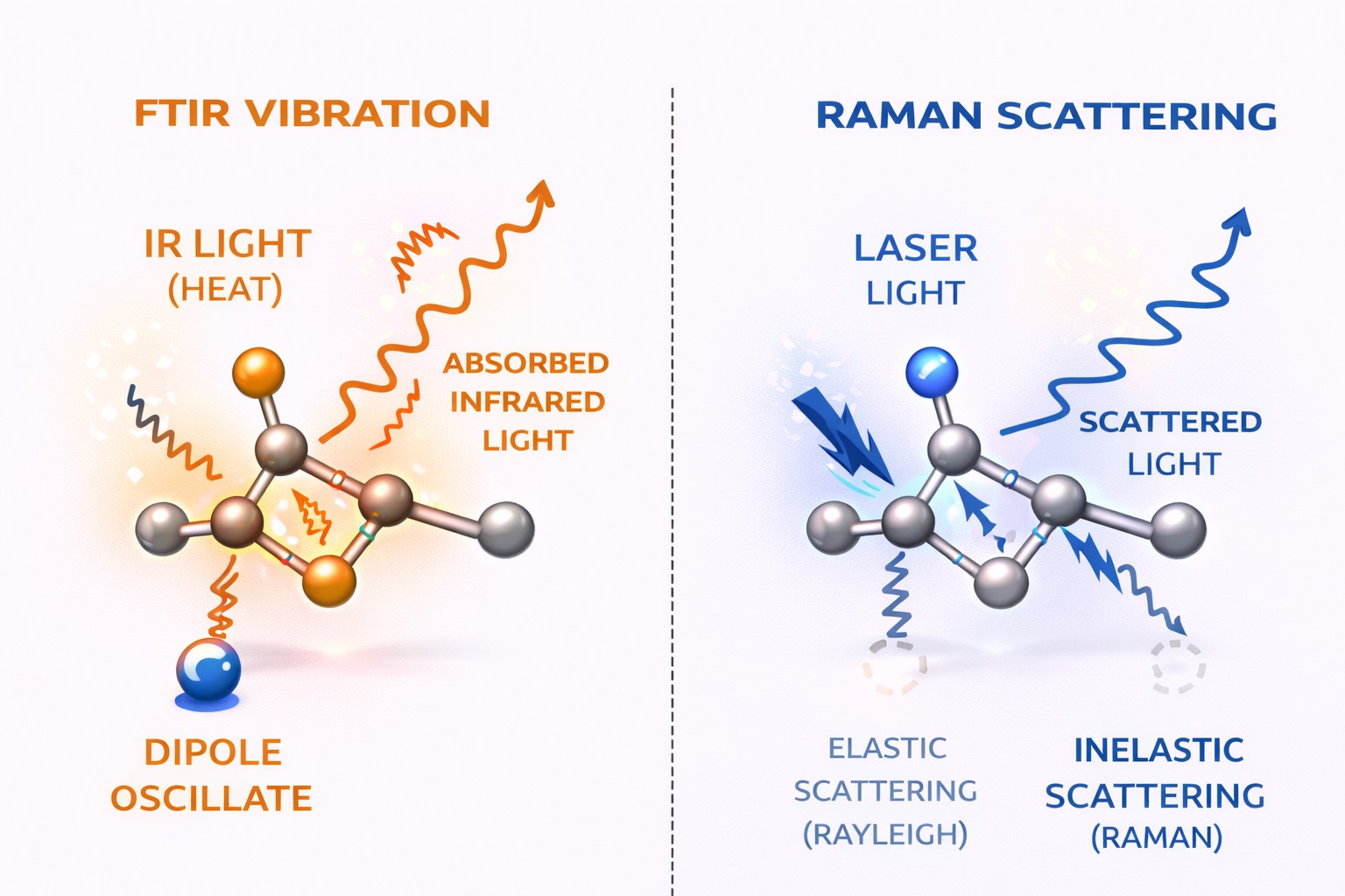
Infrared spectroscopy is based on the absorption of infrared radiation by molecular vibrations associated with a change in dipole moment, resulting in variations in bond lengths and bond angles. Raman spectroscopy, by contrast, probes molecular vibrations through inelastic scattering of incident laser light arising from changes in molecular polarizability (Figure 3). The combination of these two methods delivers complementary information on the chemical composition of materials, yielding a more accurate and complete analysis of the sample.
It is important to note that Raman and IR do exhibit complementarity for the same functional groups. Sometimes, a functional group does not show a signal in one method, underscoring the importance of applying both methods. Because most substances and functional groups are either active for Raman or infrared, AIRsight™ enables measurements with both methods – on the same spot, without moving the sample. Being active for both methods usually requires different functional groups in one molecule.
As molecular vibrational energies depend on the chemical environment, different substances exhibit characteristic infrared absorption and Raman scattering signatures. The combination of infrared and Raman spectroscopy therefore enables reliable identification and differentiation of a wide range of materials.
Figure 4 shows the layout of the IR beam path in the AIMsight™ and AIRsight™ microscopes. The sample is placed in the center, at the position enclosed by the red cones. The beam path from below enables transmission measurements, while the one from above allows reflection measurements. The wide-field camera is used to provide an overview of the area of interest. Once an interesting spot has been identified, the view can be switched to the 15× infrared objective for detailed magnification and focus adjustment. In this view, measurement points for transmission or reflection can be set with different apertures, along with an additional point for a background measurement of the surrounding air. After the background measurement, which minimizes interfering signals from CO₂ and water, the actual measurement begins with a predefined number of scans until the final spectrum appears. Following this procedure, line scans and mappings of areas of interest can also be created, consisting of multiple measurement points. The spectra obtained for each measurement point can then be compared with a database for identification.
The Raman lasers, on the other hand, are built in at the top of the microscope and can only hit the sample from the top. The 50x and 100x Raman objectives collect the backscattered light from the sample, which is then translated into a difference spectrum compared to the incident wavenumber. Therefore, Raman spectra are not measured in absolute units like FTIR but in relation to the laser light.
Putting it all together
The combination of microtome sample preparation with infrared and Raman spectroscopy allows the chemical analysis of individual layers within multilayer packaging films. By preparing thin cross-sections (Figure 5), the internal structure of such films becomes accessible for spatially resolved spectroscopic measurements. This approach is illustrated in the following example using a multilayer film taken from a commercial potato chip packaging.
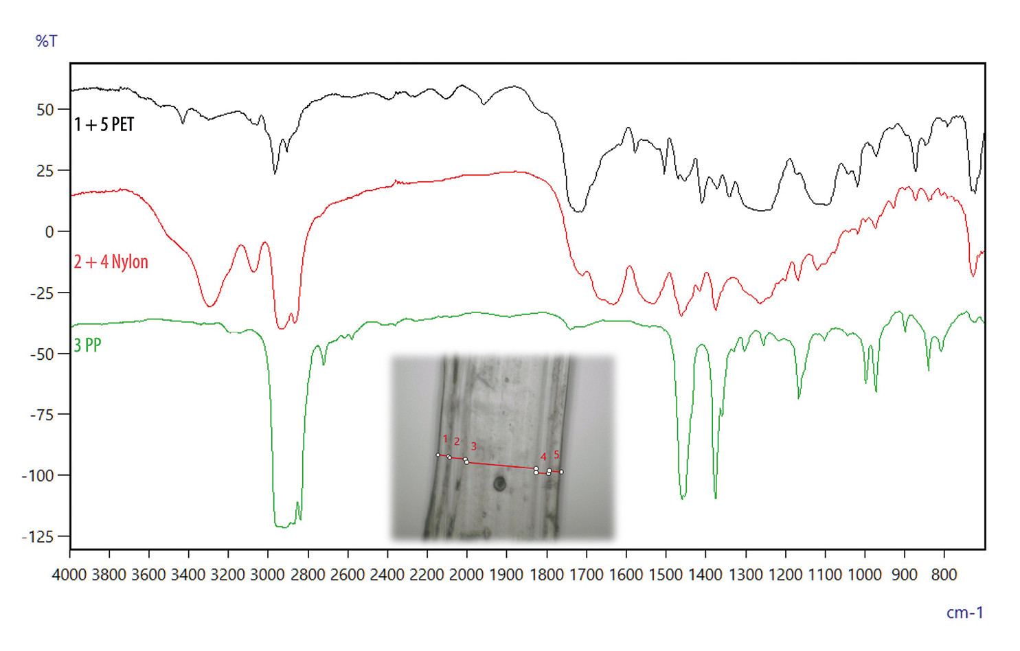
Optically, five different layers could be distinguished within the cross-section and infrared spectroscopy was used for their chemical identification. Although five individual layers were detected by Fourier transform infrared spectroscopy (FTIR) analysis, only three different materials were determined. Starting from the outer surfaces and moving toward the center of the film, the outer layers were found to consist of polyethylene terephthalate (PET), followed by layers of nylon, while the central layer was identified as polypropylene (PP) (Figure 6).
PET is commonly used as an outer layer in food packaging due to its good mechanical strength, chemical resistance and transparency, as well as its ability to provide a protective surface suitable for printing. Nylon layers are often incorporated to enhance barrier properties, particularly against oxygen and aromas, thereby contributing to extended shelf life. The central polypropylene layer offers mechanical stability and flexibility while also offering good resistance to moisture, which is essential for maintaining product quality in snack packaging.
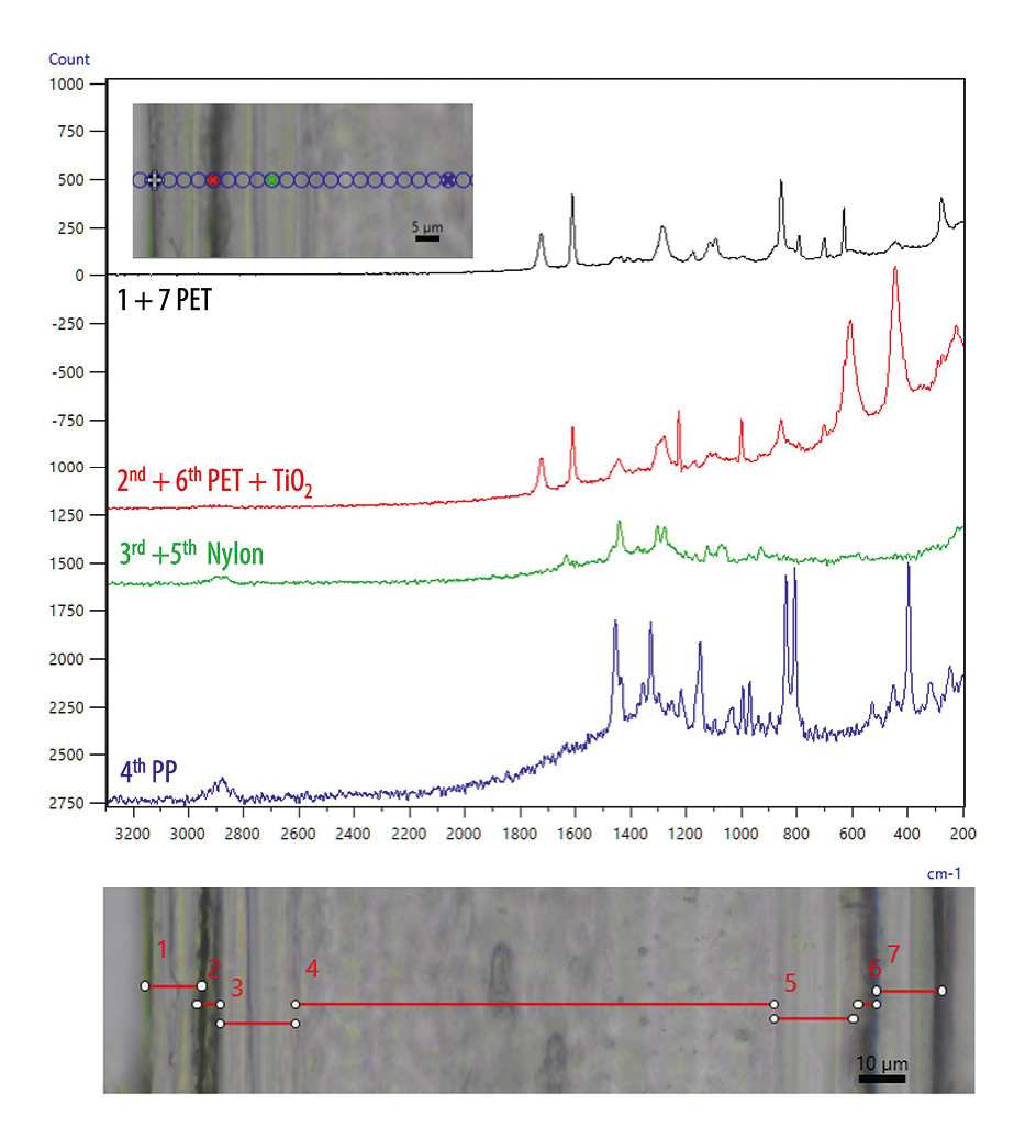
Raman spectroscopy was also employed for the analysis. In addition to the layers identified by FTIR spectroscopy, Raman analysis revealed the presence of two titanium dioxide (TiO₂) layers between the PET and nylon layers, which could not be detected using infrared spectroscopy (Figure 7). This is due to the fact that TiO₂ does not exhibit strong infrared-active vibrational modes but shows a pronounced Raman response as a result of changes in molecular polarizability. This finding highlights the advantage of combining infrared and Raman spectroscopy for the comprehensive characterization of multilayer packaging materials.
A good product requires good ingredients
Although commonly used in biological applications, it is now clear that microtome-prepared cross-sections combined with infrared and Raman microscopy can also be used to enable detailed characterization of multilayer packaging films. While infrared spectroscopy allows reliable identification of polymer layers, Raman spectroscopy offers complementary information, such as the detection of inorganic additives and precise determination of layer boundaries due to higher magnification. The analysis of a commercial chip packaging demonstrates that the AIMsight™ and AIRsight™ microscope systems are well suited for such applications, supporting quality control, material development and recycling processes in the food and pharmaceutical packaging industry. As with lasagna, putting the right things together in the right way makes all the difference.
Ščetar, M. (2021). Multilayer Packaging Materials. Packaging Materials and Processing for Food, Pharmaceuticals and Cosmetics, 131-144. Anukiruthika, T., Sethupathy, P., Wilson, A., Kashampur, K., Moses, J. A., & Anandharamakrishnan, C. (2020). Multilayer packaging: Advances in preparation techniques and emerging food applications. Comprehensive reviews in food science and food safety, 19(3), 1156-1186.Piergiovanni, L., & Limbo, S. (2016). Food packaging materials (Vol. 1671). Basel, Switzerland:: Springer.
Weblinks
Multilayer film analysis using the AIRsight™ infrared Raman microscope
A comparative study of depth and cross-section measurement of food packaging