Like putting on spectral
glasses to see hidden
colors
Like putting on spectral glasses to see hidden colors

Solving co-elution challenges for beta-blocker separation with i-PDeA II
Oktawia Kalisz and DSc Szymon Bocian, Nicolaus Copernicus University in Toruń
In liquid chromatography, separating structurally similar compounds like beta-blockers poses a challenge due to frequent co-elution. This article describes an attempt to identify eight beta-blockers within a complex mixture without laboriously injecting each compound separately. The specific goal was to find a faster, more efficient method to resolve overlapping peaks using digital tools.
In the beginning was the challenge …
Here’s the beginning of a story any researcher can relate to: A PhD student in Poland encounters a practical research challenge … In this case, the challenge was how to identify individual beta-blockers within a complex mixture in a simple and efficient way – meaning, without the need to inject each substance separately.
Clearly, she was working in the realm of high-performance liquid chromatography (HPLC), one of the most powerful analytical techniques for separating many complex mixtures of compounds. However, even with optimized chromatographic conditions, analysts frequently encounter the challenge of co-elution, where two or more compounds elute simultaneously, making their individual detection and quantification difficult. This is particularly true for structurally similar compounds, such as beta-blockers, which exhibit similar retention behaviors.

Here’s how she describes her challenge with the co-elution of beta-blockers: In a recent study, we explored the separation of eight beta-blockers using novel phosphodiester stationary phases. These were so-called polar-embedded stationary phases, which have mixed-mode properties thanks to the presence of both polar and nonpolar groups. The presence of a phosphodiester group and a hydrophobic ligand (C18 chain, C10 chain, cholesterol group or benzyl group) in the structure of the stationary phases allowed for the retention of compounds in both reversed-phase liquid chromatography (RP LC) and hydrophilic interaction liquid chromatography (HILIC) systems.
The incorporated phosphate group in this study had a pKa of 1.45; therefore a mobile phase with a pH of 7.5 was prepared. Under these conditions, both the phosphate group and the analyzed beta-blockers were ionized and so, during the analyses – in addition to hydrophobic interactions in RP LC and hydrophilic interactions in HILIC – elution occurred according to a cationic retention mechanism.
Beta-blockers were particularly interesting compounds for this study due to their various polarities and functional groups, making them ideal candidates to characterize the selectivity of four novel materials (Diol-P-C10, Diol-P-benzyl, Diol-P-chol and Diol-P-C18). Initially, we attempted to separate the beta-blockers using isocratic and gradient elution. However, this approach was insufficient to achieve complete resolution of all compounds in the mixture due to peak co-elution.
In the PhD student’s case, the challenge became clear from the very first trials on novel phosphodiester stationary phases. Instead of neat, sharp peaks, the chromatograms showed tailing and broad peaks, while several compounds co-eluted into a single signal. Adjusting gradients and modifying the mobile phase brought improvement but it was not sufficient. Some analytes continued to remain hidden under overlapping peaks. What to do?
Looking at the problem from another angle
At this point, the PhD student did what anyone might do: She talked with her advisor, who suggested a different approach. “You don’t have to strive for perfect separation if it’s not necessary,” advised Dr. Szymon Bocian (Nicolaus Copernicus University, Toruń, Poland). “Let’s look at the data from a different perspective. Try i-PDeA II.”
i-PDeA II (Intelligent Peak Deconvolution Analysis II) is an advanced data processing technique available in the LabSolutions software. Dr. Bocian was already familiar with i-PDeA II through previous collaboration with Shimadzu. He also knew the capabilities of PDA detectors, which capture full UV spectra across the chromatogram. That knowledge led him to suggest a new strategy: Instead of endlessly optimizing the chromatographic conditions, why not use spectral deconvolution to reveal what the chromatogram could not show?
Why not use digital peak separation?
While the phosphodiester stationary phases and controlled mobile phase pH had provided improved chromatographic performance, some beta-blockers had still exhibited significant co-elution. Instead of resorting to extensive method development to achieve baseline separation, the Toruń team now began to use the digital i-PDeA II tool to deconvolute overlapping peaks directly from the acquired chromatographic data. This tool, in combination with photodiode array (PDA) detectors, enabled the use of 3D PDA data to mathematically differentiate overlapping peaks, even if they were not visually separated on the chromatogram. (This method applies the mathematical derivative of a spectrum to distinguish overlapping signals. Since the first derivative becomes zero at maxima and minima, individual peaks can be identified, and the chromatograms of each component can be created.)
Instead of relying solely on retention time, i-PDeA II was able to analyze the entire UV-Vis spectral profile on the chromatogram and extract the spectra of the pure compounds that were components of the mixture. This approach not only provided better identification, it also minimized both method development and analysis time. In the case of the PhD student’s research, it was a turning point.


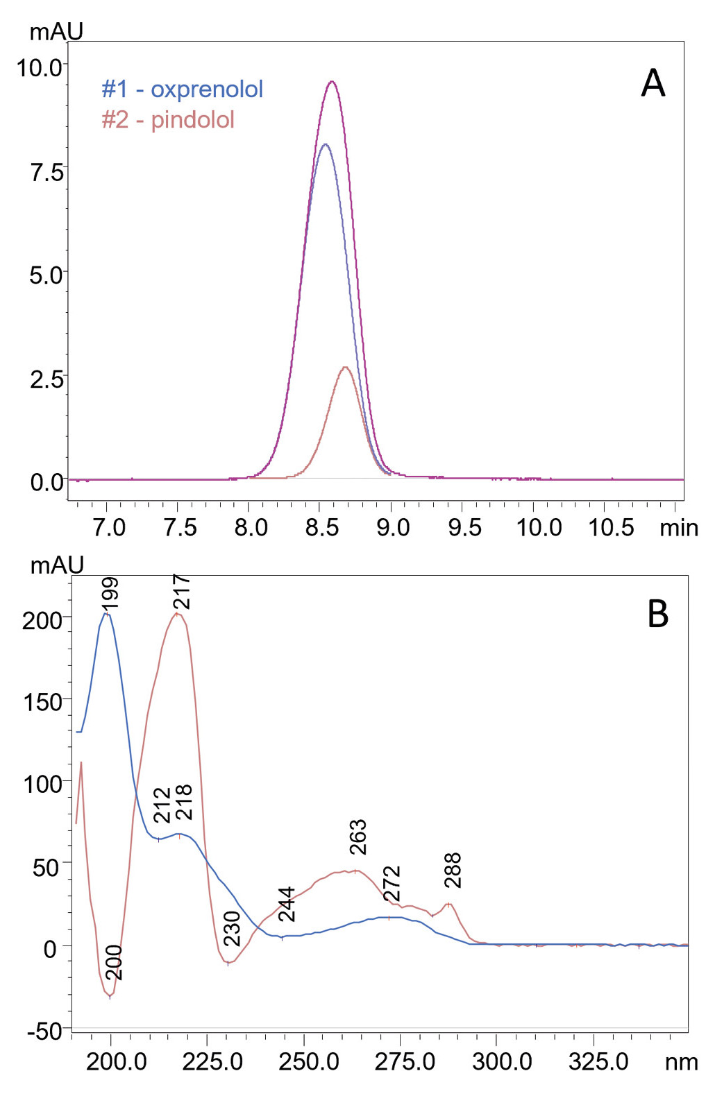
Success no. 1: Resolving oxprenolol and pindolol
One of the most significant challenges the team faced was the co-elution of oxprenolol and pindolol on a Diol-P-C10 stationary phase under HILIC conditions. These compounds had a selectivity factor of only 1.01, meaning their peaks were nearly indistinguishable. Without deconvolution, the detector recorded a single peak, making individual analysis impossible. However, i-PDeA II successfully separated the two components digitally by extracting their individual spectra (see Figure 1 and Table 1).
| Column | Diol-P-C10 (125 mm 4.6 mm, 5 µm) |
| Mobile phase | 10 mM CH3COONH4 in H2O pH 7.5:ACN (10:90 v/v) |
| Flow rate | 1 mL/min |
| Temperature | 25°C |
| Injection volume | 10 µL |
| Analytical wavelength | 254 nm |
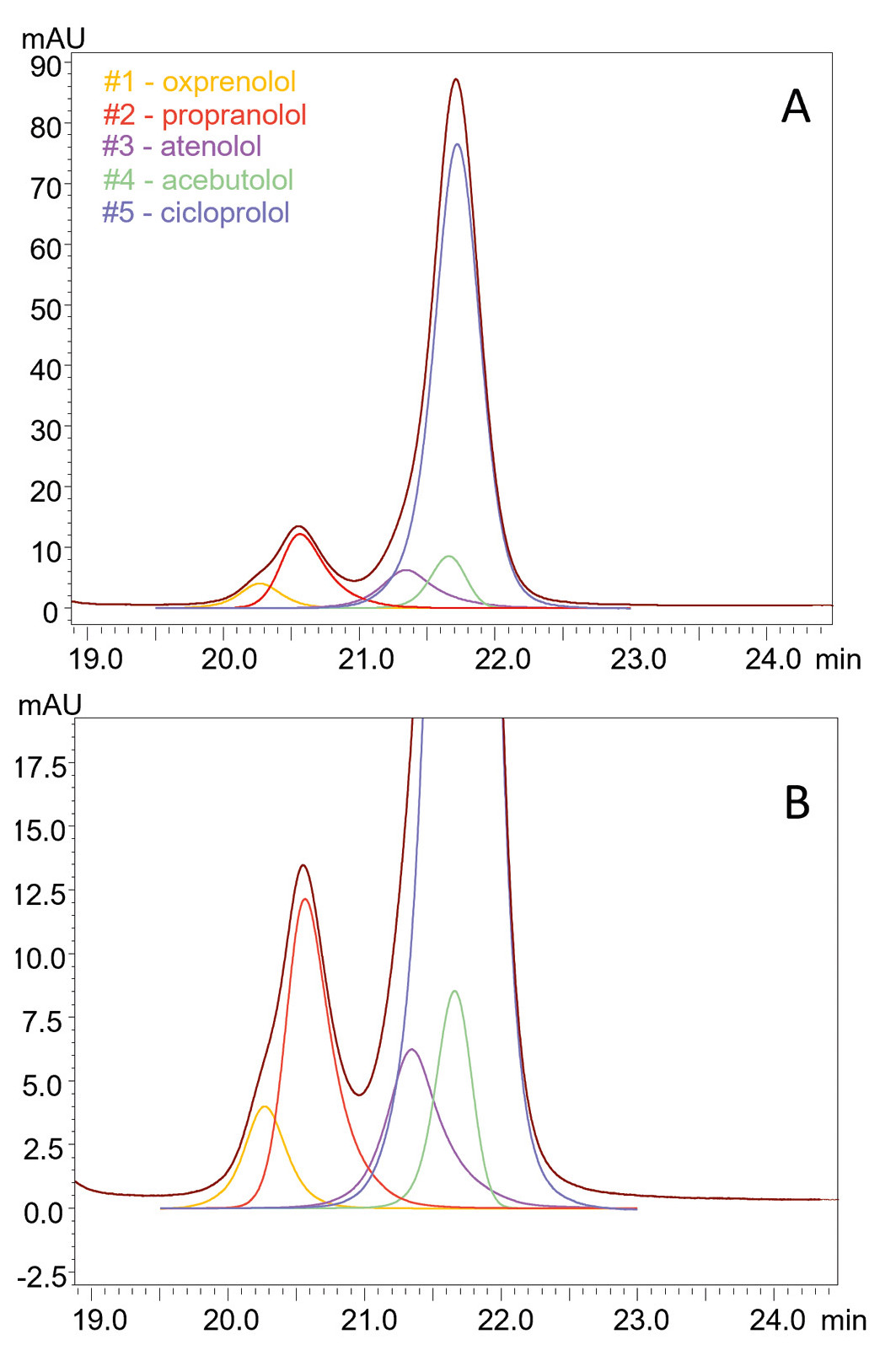
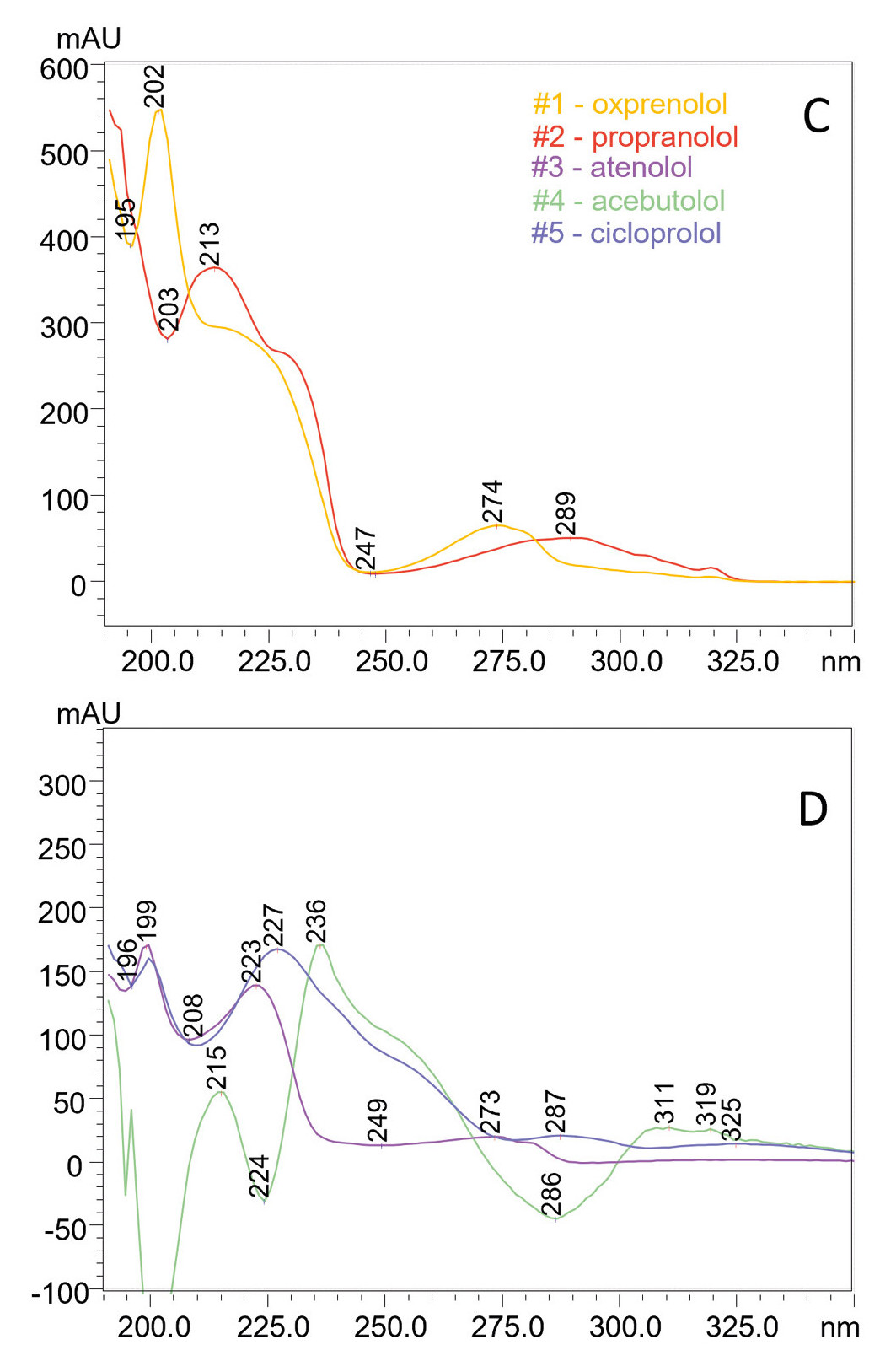
Success no. 2: Simultaneous deconvolution of five beta-blockers
The application of i-PDeA II also demonstrated that this function is not only effective for resolving two peaks (such as distinguishing an impurity from the main analyte) but also for deconvoluting multiple co-eluted compounds from complex mixtures. In a gradient separation performed on the Diol-P-benzyl stationary phase, five beta blockers – oxprenolol, propranolol, atenolol, acebutolol and cicloprolol – were co-eluted. The i-PDeA II software analyzed the UV spectral differences of each compound, which allowed their identification (see Figure 2, Figure 3 and Table 2).
When traditional separation fails, i-PDeA II succeeds
The PhD student’s challenge was that standard HPLC methods could not adequately separate beta-blockers and address the co-elution of structurally similar compounds. This prompted her to look for an innovative solution, which she – with the help of her advisor – found in the integration of i-PDeA II with phosphodiester stationary phases and which were used in this way for the first time in her study.
This integration proved to be an indispensable tool in this study of beta-blocker separation. The digital separation technique allowed the researchers to resolve co-eluted peaks based on their unique UV spectra, significantly improving the reliability of analysis. The results demonstrated that deconvolution can replace the need for extensive method development and considerably reduce analysis time while maintaining high specificity.
| Column | Diol-P-benzyl (125 mm 4.6 mm, 5 µm) |
| Mobile phase (A: 10 mM CH3COONH4 in H2O pH 7.5; B: ACN) |
Gradient elution: 0 min – 11.8 min: 86% B 11.8 min – 11.9 min: 59% B |
| Flow rate | 1 mL/min |
| Temperature | 25°C |
| Injection volume | 10 µL |
| Analytical wavelength | 254 nm |


A clear step forward in chromatographic analysis
Among pharmacologically active substances, beta-blockers are a group of drugs used in cardiology. They are also administered to animals during transport to reduce their morbidity, which consequently results in their presence in meat and other animal products. The method developed here provides a beneficial new way to analyze many reference and real samples (blood, urine or wastewater) used in drug metabolism studies, anti-doping tests and trace analysis.
These findings also highlight the broader applicability of i-PDeA II, showcasing its potential as a powerful analytical tool for resolving co-eluting compounds in complex mixtures. This methodology can also be particularly useful in drug quality control for faster and more accurate detection of impurities that may be hidden under the peak of the active substance.
Or, as the PhD student puts it, “Using i-PDeA II reveals hidden details invisible to the naked eye in traditional LC. It is like putting on spectral glasses that allow you to see individual colors in a beam of light that otherwise appears white.”
1. Kalisz O., Dembek M., Studzińska S., Bocian S. (2023). Beta-Blocker Separation on Phosphodiester Stationary Phases – The Application of Intelligent Peak Deconvolution Analysis. Molecules. 28: 3249. doi: 10.3390/molecules28073249.
2. Vecchietti D., Nishio A., Fujita Y., Yoshida T., Yanagisawa T., Kou D. (2022). Liquid chromatography coupled with photodiode array and a multivariate curve resolution – Alternating least square algorithm for identification and quantification of co-eluting impurities in pharmaceutical analysis. J Chromatogr A. 1678: 463364. doi: 10.1016/j.chroma.2022.463364.